Data Samples
Data samples of the various modalities used to monitor patients:
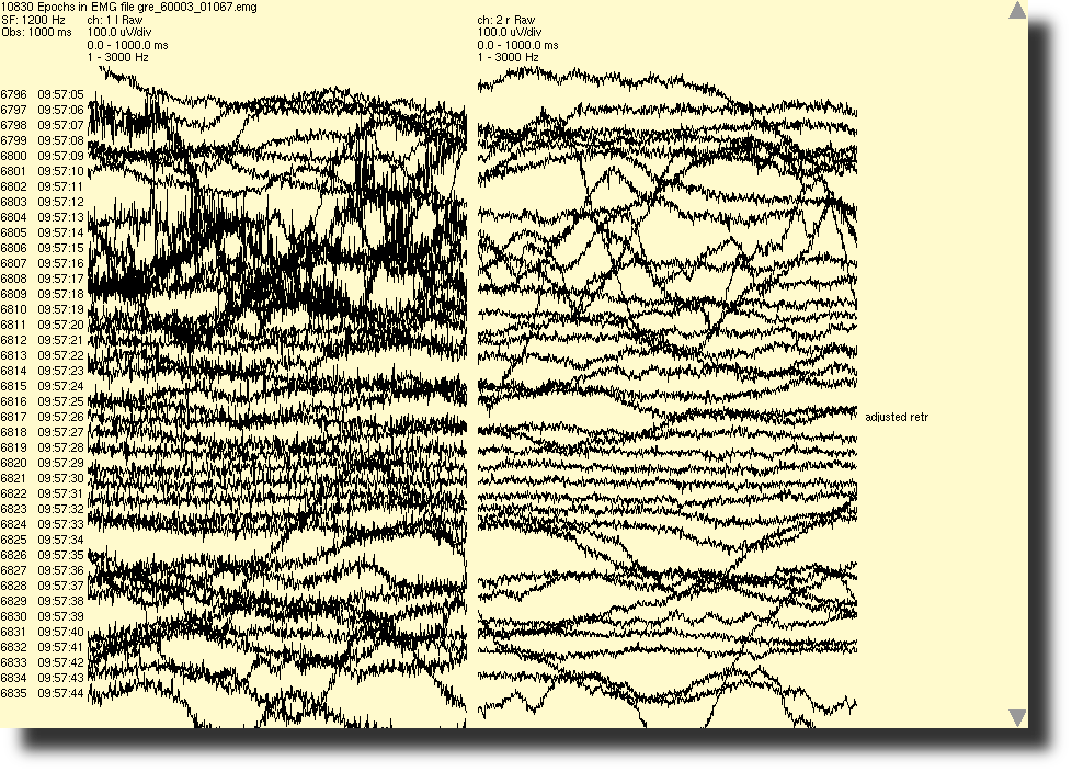
Below is an example irritation of spontaneous EMG of the left and right recurrent laryngeal nerve being monitored during a thyroidectomy.
Click Image for Enlargement
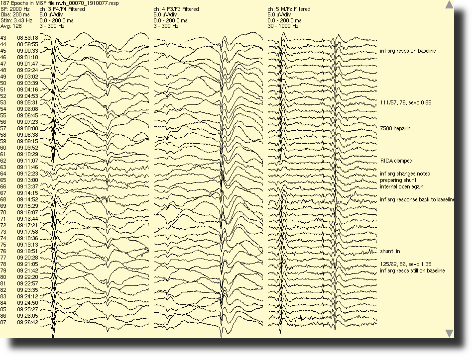
Shown below is SEP data of the median nerve (MSP) observed during a carotid endarterectomy. Changes can be seen in the responses from the left median nerve at the time of clamping of the common carotid artery.
Click Image for Enlargement
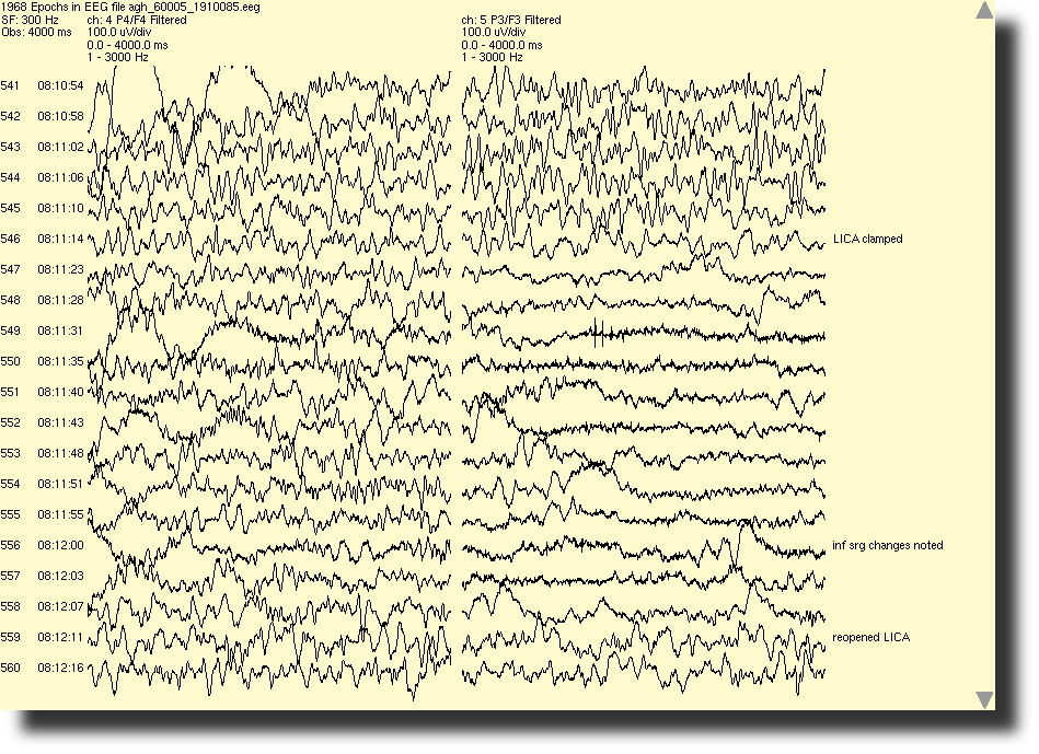
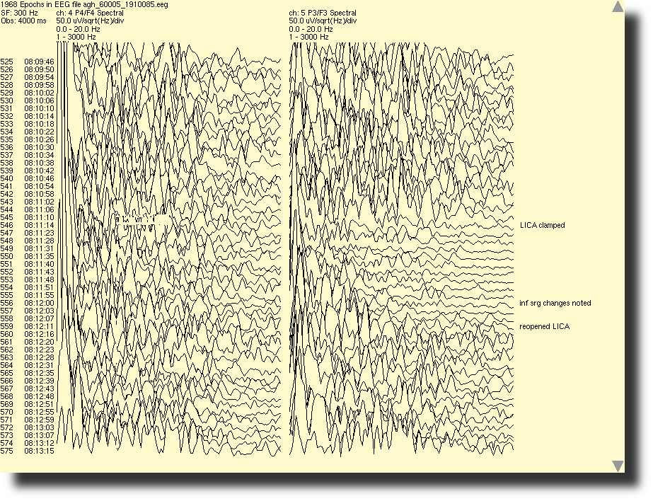
An example of filtered EEG (left) and spectral EEG (right) during a carotid endarterectomy is pictured here. A decrease in cortical activity in the left hemisphere occured when the left common carotid artery was clamped during the procedure.
Click Images for Enlargement
Below is sample data of MEP collected during a craniotomy (left) and a shoulder arthroplasty (right) where the motor cortex is stimulated and potentials are recorded from muscle groups.
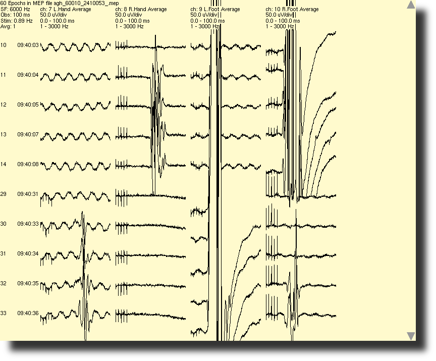

Click Images for Enlargement
Below is a sample of CMP data from a posterior lumbar fusion. As the pedicle screws are stimulated, evoked responses are obtained in several lower extremity muscle groups.
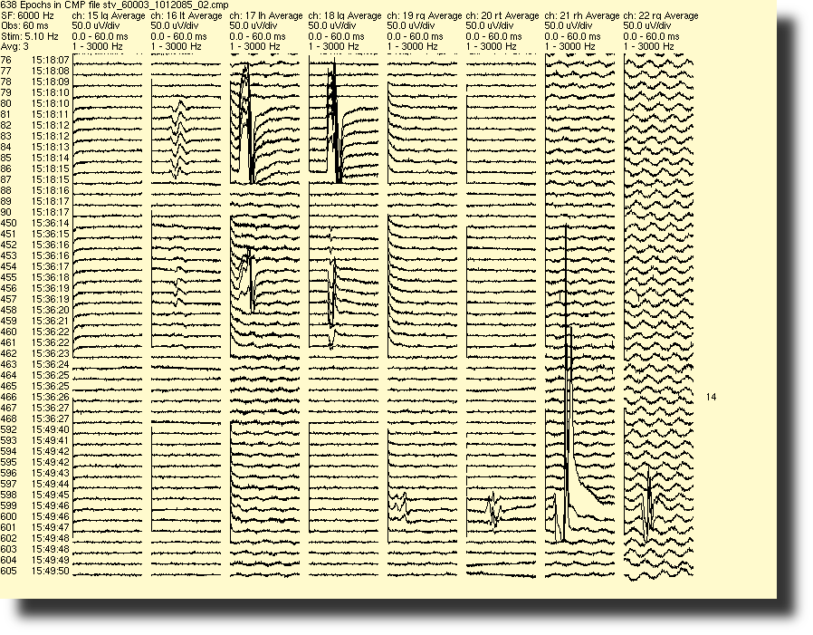
Click Image for Enlargement
This is an example of data collected from a BAEP test monitoring CN VIII, during a retromastoid craniotomy for microvascular decompression of the trigeminal nerve.
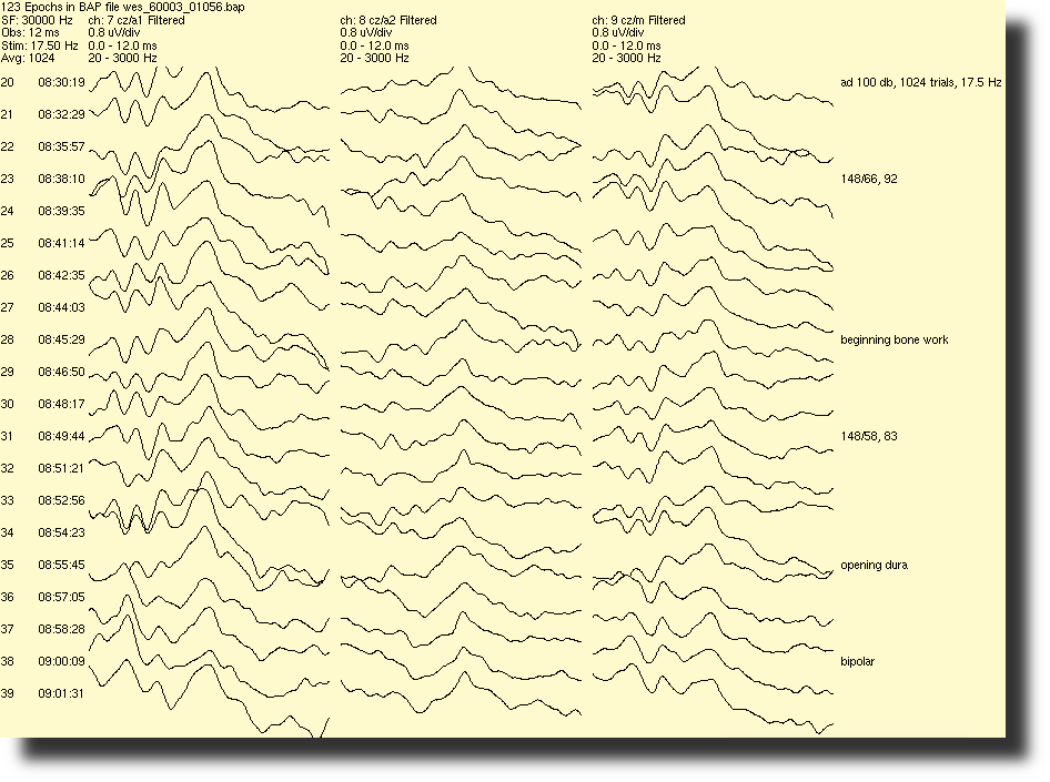
Click Image for Enlargement